|
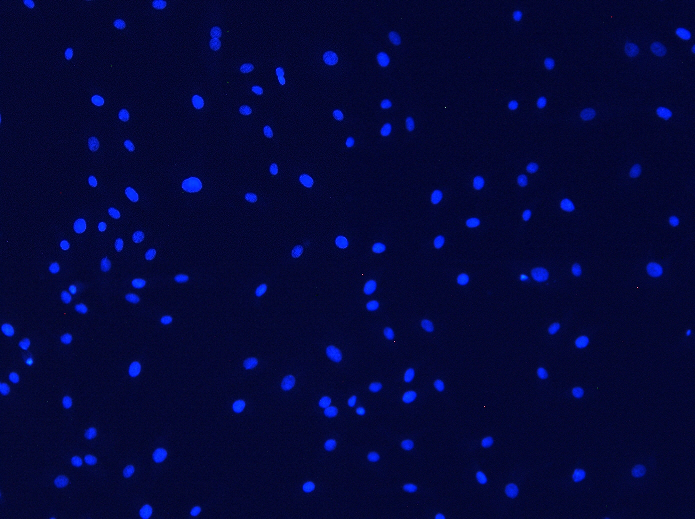
Objective 10X. Exposure time 800 ms. |
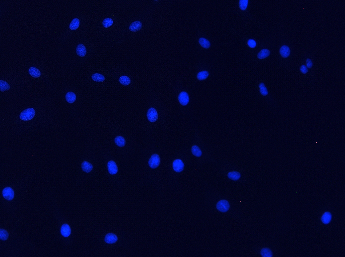
Objective 20X. Exposure time 400 ms. |
Home > LED Light Sources > Images and Exposure Times with 030-365 Light Source
Images and Exposure Times with TOFRA 030-365 LED Light Source
Tests were performed on Olympus BX51 microscope with 10X UPlanFL 0.3NA and 20X UPlanFL 0.5NA objectives and Chroma 39000 - AT - DAPI/Hoechst/AlexaFluor 350 filter cube.
The light source was set to full intensity. Focusing of the light source was done by focusing the LED chip on the specimen without objective in the optical path. The specimen (fluorescent plastic) was pre-focused with a 10X objective.
Camera - Basler color camera scA 1400-17gc, chip size 1390*1038 pixels, pixel size 6.45 μm * 6.45 μm, in 8-bit mode, Gain 600. Images in the table are full camera frames scaled down by a factor of 2. Exposure time was chosen to slightly saturate the image. No digital enhancement was done.
Specimens: Molecular Probes FluoCells Prepared Slide #1- bovine pulmonary artery endothelial (BPAE) cells with MitoTracker Red CMXRos, Alexa Fluor 488 Phalloidin, and DAPI.
|
Objective 10X. Exposure time 800 ms. |
Objective 20X. Exposure time 400 ms. |